New Study Finds Tumor Volume Doubling Time May Predict Progression Risk in Papillary Thyroid Cancer
In the absence of biological or molecular predictive markers, tumor volume doubling time is a good indicator for predicting the growth rate of papillary thyroid carcinomas during active surveillance, according to findings from a single-center cohort study recently published in <em>Thyroid</em>.


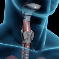
thyroid cancer
In the absence of biological or molecular predictive markers, tumor volume doubling time (TVDT) is a good indicator for predicting the growth rate of papillary thyroid carcinomas (PTC) during active surveillance, according to findings from a single-center cohort study recently published inThyroid.1
Among 273 patients, 10.3% of the patients had TVDT < 2 years, 5.1% had TVDT 2 to 3 years, 6.2% had TVDT 3 to 4 years, 6.6% had TVDT 4 to 5 years, and 71.8% had TVDT ≥ 5 years. Patients were classified into 2 groups: the rapid-growing group (TVDT < 5 years) and the stable group (TVDT ≥ 5 years). Notably, patients in the rapid-growing group (77 patients, 28.2%) were significantly younger than those in the stable group (196 patients, 71.8%;P=0.004).
“Younger age and macrocalcification in the initial ultrasound were associated with rapid-growing PTCs,” wrote the authors, led by Hye-Seon Oh, MD, of the National Police Hospital in Seoul, Korea. “Determination of TVDT during the early phase of active surveillance may be helpful for the prediction of rapidly progressing PTCs and prompt an early decision for a surgical approach.”
At diagnosis, patients received ultrasonography that documented maximum tumor diameter, volume, location, contents, echogenicity of solid portion, shape, margin, calcifications, and subcapsular location of the tumor.2
Exclusion factors included lateral cervical lymph node metastasis, macroscopic invasion into perithyroidal soft tissue, or invasion into the trachea. The investigators also excluded patients with distant metastasis or diagnosis with an aggressive variant of papillary thyroid cancer.
Study participants received follow-up physical examinations and neck ultrasound every 6 to12 months. The investigators recommended thyroid surgery when the primary tumor was larger than 1 cm, increased by more than 3 mm, or tumor volume increased by more than 50% from the baseline. They also recommended thyroid surgery when new metastatic lymph nodes or distant metastasis were found or when the tumor grew toward adjacent structures.
Approximately three-quarters of the patients were female. Their median age at diagnosis was 51.1 years (Interquartile Range [IQR] 42.261.0), and 115 patients (42.1%) were younger than 50 years.
Patients’ initial maximum tumor diameter was 5.8 mm (IQR 4.47.3), while the initial maximum tumor volume was 62.1 mm3(IQR 28.1122.8). Median patient follow-up was 42 months (IQR 28.6–60.8). During follow-up, nearly one-fourth of patients experienced a tumor volume increase ≥ 50% and 12 patients (4.4%) had significant maximal diameter increase ≥ 3 mm.
Oh et al found no difference in TVDT based on patient sex, initial maximal diameter, or initial volume. They did find, however, that patients’ ages did correspond to different TVDTs (P= 0.037). They found a maximal diameter increase ≥3 mm only in patients with TVDT < 2 years (n = 9) and 2 to 3 years (n = 3).
In examining differences between the stable and high-risk groups, the investigators found no significant differences in sex, initial maximal tumor diameter or initial tumor volume. However, the tumor volume in the high-risk group increased over time (P= 0.008), while the tumor volume in the stable-disease group remained steady (P<0.001).
They also found significant sonographic differences between the 2 groups, specifically in terms of calcification pattern (P= 0.001). Nearly a quarter (23.4%) of the fast-growing group had macrocalcification, while only 8.2% of the stable group did. In the high-risk group, only 1 patient displayed calcified spot and rim calcifications. They found no significant differences between the groups in terms of tumor location, internal content, echogenicity, shape, margin, and subcapsular location.
Multivariate analysis found that age younger than 50 was significantly associated with rapid tumor growth of PTC (Odds Ratio [OR], 2.31; 95% confidence interval (CI), 1.304.31;P= 0.004]. Likewise, macrocalcification found at initial ultrasound was independently associated with rapid tumor growth of PTC (OR, 4.98; 95% CI, 2.1911.69;P< 0.001).
During follow-up, 53 patients (19.4%) required thyroid surgery, with a median time from diagnosis of 28.6 months (IQR, 21.740.2). Of these patients, 32 (60.3%) were in the rapid-growing, high-risk group. Final pathology reports indicated that 17 patients (32.1%) had lymph node metastasis. Two patients in the high-risk group had lateral cervical lymph node metastasis. During a median postsurgical follow-up period of 14.0 months (IQR, 4.8–24.3), no patients experienced recurrences, distant metastasis, or cancer-specific death.
Oh et al concluded that TVDT is a good indicator to predict how quickly papillary thyroid carcinomas might grow during active surveillance and could prompt an early decision for a surgical approach. “Determination of TVDT in active surveillance is advantageous because it reflects the time-dependent growth rate of PTCs. This approach of TVDT is not consistent with the current definition of significant increase of maximal diameter and relative TV increase,” they wrote. “However, all patients with a maximal tumor size increase ≥ 3 mm had TVDT < 3 years and 89.8% of patients with volume increase ≥ 50% had TVDT < 5 years.”
In noting study limitations, Oh et al listed its single-center and retrospective status and relatively small size, adding that selection bias was possible. Given the natural history of papillary thyroid cancer during active surveillance, they suggest conducting longer-term prospective studies.
References:
- Oh HS, Kwon H, Song E, et al. Tumor Volume Doubling Time in Active Surveillance of Papillary Thyroid Carcinoma.Thyroid2019. DOI: 10.1089/thy.2018.0609 Epub ahead of print.
- Oh HS, Kwon H, Song E, et al. 2018 Preoperative Clinical and Sonographic Predictors for Lateral Cervical Lymph Node Metastases in Sporadic Medullary Thyroid Carcinoma.Thyroid2018; 28:362-368.


Anticipating Novel Options for the RAI-Refractory DTC Armamentarium
May 15th 2023In season 4, episode 6 of Targeted Talks, Warren Swegal, MD, takes a multidisciplinary look at the RAI-refractory differentiated thyroid cancer treatment landscape, including the research behind 2 promising systemic therapy options.
Listen
Optimizing Thyroid Cancer Care: New Tools and Emerging Options
March 12th 2024Lori J. Wirth, MD, discussed advancements in diagnosing and treating thyroid cancer, offering community oncologists improved tools for identifying low-risk cancers, personalizing treatment, and exploring innovative approaches.
Read More

