Now Playing
Hope Rugo, MD, FASCO: Welcome to this Targeted Oncology “Breast Cancer” Virtual Tumor Board®. Thank you for participating. Today, we’re going to talk about a number of different cases that I think will be pertinent to your clinical practices regarding new ways of treating breast cancer. Our participants today include a number of medical oncologists as well as a pathologist.
I’m Dr Hope Rugo, and I’m joined by Evita Sadimin, who is a pathologist at the Rutgers Cancer Institute of New Jersey and has a lot of experience with decision-making and testing for HER2-positive breast cancer, PD-L1.
I’m also joined by 3 medical oncologists: My colleagues Dr Adam Brufsky, from the University of Pittsburgh Medical Center; Dr Ian Krop, from Dana-Farber Cancer Institute; and Dr Tiffany Traina, from Memorial Sloan Kettering Cancer Center.
I'm Dr Hope Rugo, from the University of California, San Francisco Comprehensive Cancer Center.
Today, as I mentioned, we’re going to focus on breast cancer. My colleagues and I will review 4 clinical cases. We’ll discuss an individualized approach to treatment for each patient, and we’ll review key clinical data that impacts our decision. Then, the panel will discuss how these data impact clinical practice in each setting. Thank you for joining us. Let’s get started with our first case.
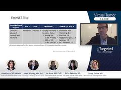
Dr Ian Krop will talk about a case of a patient with early-stage HER2-positive breast cancer.
Ian Krop, MD, PhD: Thanks, Hope. I think I have a case that’s interesting, both because it presents some good medical oncology discussion points but also because it presents some good pathology discussion points.
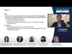
This is a 52-year-old postmenopausal woman who presented with palpable masses in her breasts and axilla on a routine annual exam. This is an otherwise healthy woman. She has 2 children. Age of menopause was 49. She doesn’t have any significant comorbidities and has no family history of cancer.
On that initial imaging to work up the palpable mass, a mammogram demonstrated a 2.7-cm tumor in her right breast, corresponding to the palpable abnormality, and a breast MRI confirmed the tumor as well as right axillary lymph node involvement with an approximately 2.5-cm node. This led to a biopsy of both the breast and the node. The biopsy of the breast demonstrated poorly differentiated invasive ductal carcinoma. The cancer was strongly estrogen receptor–positive in 90% of cells, progesterone receptor–negative, and there was a relatively high Ki-67 expression of 50%.
The HER2 analysis showed that the IHC [immunohistochemistry] was 2+, or equivocal, but the follow-up FISH [fluorescence in situ hybridization] demonstrated 4.5 copies of HER2 and a HER2-to-CEP17ratioof 2.2. So, this was a positive FISH result. The FNA [fine needle aspiration] of the axillary node confirmed adenocarcinoma in the node.
Based on these results, she went on to receive neoadjuvant systemic therapy with dose-dense AC [doxorubicin and cyclophosphamide] followed by paclitaxel with trastuzumab and pertuzumab. With the first cycle, she did fine. But in the second cycle, she developed grade 3 diarrhea. We instituted an aggressive anti-diarrheal regimen with loperamide, and the pertuzumab was held for that cycle.
Fortunately, the diarrhea then resolved, and she was able to complete the remaining cycles of paclitaxel with trastuzumab and pertuzumab. She then underwent breast-conserving surgery and an axillary dissection and, fortunately, was found to have a pathologic complete response.
Transcript edited for clarity.