Diagnosis of Metastatic GIST
Jonathan Trent, MD, PhD: A 64-year-old Caucasian male presents to the emergency room with fatigue and abdominal pain. He has no significant past medical history other than well-controlled hypertension that is managed with a beta blocker; he has no significant family history. An abdominal CT scan is performed and this reveals a 12-cm gastric mass involving the cardia and fundus, as well as a 7-cm solitary liver tumor.
A biopsy was performed revealing a gastrointestinal stromal tumor, or GIST, that was KIT-positive by immunohistochemistry, and found to have anexon 9mutation. The tumor was also found to be high-grade, with a greater than 5 per 50 high-power field mitotic rate. The patient was initiated on therapy with imatinib at the 600 mg per day dose; this was well toleratedthe patient maintained therapy for 5 months. At that time, the patient had another CT scan performed of the abdomen that revealed decrease in size of the solitary liver metastasis from 7 cm to 4 cm, with relative stability of the gastric mass. The patient was referred to a surgical oncologist who performed an R0 resection with negative margins of both the liver metastasis and the primary tumor in the stomach. Postoperatively, the patient recovered and was placed on imatinib 800 mg per day.
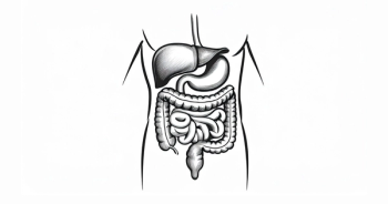
Patients with gastrointestinal stromal tumors present with a variety of different symptoms, largely depending on the location of the primary tumor. This patient had a primary tumor in the stomach. The patient presented with fatigue, probably related to anemia, as well as abdominal painthis presentation is consistent with a gastrointestinal stromal tumor in this anatomic location.
This patient with GIST was initially treated with 600 mg per day of imatinib, underwent surgical resection and postoperatively was placed on 800 mg per day of imatinib. This management is supported by the GIST metaanalysis that found that patients who have anexon 9mutation benefit from higher doses than 400 mg per day of imatinib, in terms of not only progression-free survival, but overall survival as well.
Transcript edited for clarity.
September 2014
- A 64-year old Caucasian male presented with abdominal pain and 3-month history of fatigue
- PMH was remarkable for hypertension well-controlled with a beta-blocker
- No family history of cancer
- He could perform all activities independently
- Abdominal CT findings:
- 12-cm mass arising from the stomach and involving the cardia, fundus, and body of the stomach
- 7-cm solitary mass in the left lobe of the liver
- Biopsy results:
- Gastric GIST with liver metastases
- IHC positive for CD117 (c-KIT), molecular analysis showed exon 9 deletion
- Mitotic activity, high with >5 mitoses/50 HPFs
- Treatment was initiated with neoadjuvant imatinib 600 mg daily for 5 months
- The primary tumor was stable during this time, the liver mass size decreased from 7 cm to 4 cm
- The patient was referred to a surgeon and underwent hepatectomy for the liver metastasis
- Following surgery, R0 resection with clear margins
- Treatment was initiated with imatinib 800 mg daily
August 2016
- Abdominal CT imaging findings:
- Multiple peritoneal implants
- A new small nodule (<1 cm) in the liver
- The patient could perform all activities independently with small occasional breaks, but could not perform physically strenuous activities
- He was switched to sunitinib 37.5 mg daily
February 2017
- At his 6-month follow-up, the patient was still able to perform most non-strenuous activities independently; however, the frequency of being able to do so had declined significantly
- Abdominal CT scan showed progression in multiple peritoneal implants; the liver nodule increased in size to 2 cm
- He was referred to an academic center
- His treatment was switched to regorafenib 160 mg, 3 weeks on, 1 week off
- The patient appeared to tolerate therapy well, after initial dose modification due to diarrhea experienced during the second week of therapy
- At the 6-month follow-up:
- Abdominal CT scan showed slight reduction in the peritoneal implants
- The liver nodule was no longer visible


