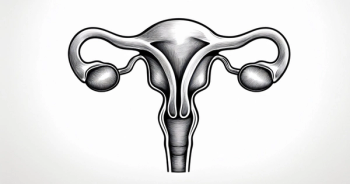
Case 1: Stage IV Ovarian Cancer With Multiple Metastatic Sites
EXPERT PERSPECTIVE VIRTUAL TUMOR BOARD
Robert L. Coleman, MD:Thank you for joining us for thisTargeted Oncology Virtual Tumor Board®, which is focused on ovarian cancer. In today’sTargeted Oncology Virtual Tumor Board®presentation, my colleagues and I will review 4 clinical cases. We will discuss an individualized approach to treatment for each patient and will review key trial data that impact our decisions.
I am Dr Robert Coleman, professor in the Department of Gynecologic Oncology and Reproductive Medicine, and executive director of MD Anderson Cancer Network Research, and the Ann Rife Cox chair in gynecology at The University of Texas MD Anderson Cancer Center in Houston, Texas.
Today I’m joined by Dr Susana M. Campos, assistant professor of medicine in the Division of Medical Oncology at Dana-Farber Cancer Institute at Harvard Medical School in Boston, Massachusetts;
Dr David O’Malley, medical director of gynecologic oncology, director of clinical research of gynecologic oncology, co-director in the Gynecologic Oncology Phase I Program, and professor at The Ohio State University and the James Cancer Center in Columbus, Ohio;
Dr Gregory Riedlinger, assistant professor of pathology in the Division of Translational Pathology at Rutgers Cancer Institute of New Jersey and Rutgers Robert Wood Johnson Medical School in New Brunswick, New Jersey;
and Dr Shannon N. Westin, associate professor and director of early drug development in the Department of Gynecologic Oncology and Reproductive Medicine at The University of Texas MD Anderson Cancer Center in Houston, Texas.
Thank you for joining us. Let’s get started with our first case.
Our first case will be discussed by Dr Susana Campos. Why don’t you take it away, Susana?
Susana M. Campos, MD, MPH:Thanks. I’d like to share with you guys a case that’s quite interesting. It’s a 44-year-old premenopausal woman who presented to her gynecologist complaining of weakness and vague abdominal discomfort for a duration of about 4 months. Her past medical history is mainly unremarkable. She has 2 children and has always had a normal menstrual cycle. In terms of her family history, both of her parents are living. There is a history of lung cancer on her father’s side. On a physical examination, what was dominant was really right-sided abdominal tenderness and swelling. Imaging studies were done, which showed a right-sided solid cystic adnexal mass, measuring about 8.8 centimeters in size. There was periaortic lymphadenopathy, omental caking, a solitary liver lesion, and a splenic mass. A percutaneous biopsy did show high-grade ovarian adenocarcinoma.
Robert L. Coleman, MD:OK, so it was a good case. It is one we see frequently. First, talk a little about the pathology. Give us your thoughts on what you are thinking about this pathology.
Gregory Riedlinger, MD, PhD.:Particularly as a pathologist, we’re just looking at tissue initially on an H&E [hematoxylin and eosin stain] slide. There are certain morphologic features for which we’re thinking about, depending on the specific diseases. In this case, they’re saying high-grade ovarian cancer. So we’re usually looking for more of a serous morphology. And then it’s an adenocarcinoma, so it’s a gland-forming tumor. The pathologist is not going any further than that without immunohistochemistry. So you’re going to be flying a bit blind.
So typically, a pathologist will use a panel markera PAX8 transcription factor—to kind of confirm that it’s of Mullerian origin. And then, with the high-grade serous, they tend to uniformly actually have a p53 mutation. So on immunohistochemistry, there are actually 2 phenotypes. There are the high expressors, where all of the tumor cells would have p53; or the null phenotype, where none of the cells have p53. And actually, when we do further molecular testing, this correlates with the actual underlying p53 mutation we see. With theTP53missense mutations, these are always the high expressors that actually stabilize the p53 protein. Whereas the p53 frame shifter truncating correlate with loss of function. And so, that’s kind of consistent with the high-grade serous.
In ovarian, a lot of times we’ll use a marker like WT1, and that’s kind of what tends to be seen a little bit more somewhere in the peritoneum, indicating a peritoneal origin. But there is kind of extensive literature now on what’s called high-grade serous ovarian carcinoma really originating in the fallopian tubes. So women had been getting prophylactic BSOs [bilateral salpingo-oophorectomies], who were germlineBRCA1andBRCA2carriers. And a number of groups set up protocols to really section through the end of the fallopian tube and looked for lesions there. They found these serous tubal intraepithelial carcinomas [STICs], which were believed to potentially be the origin for these ovarian carcinomas.
And then, work came out inNature Communications. Even before the serous tubal intraepithelial carcinoma, which would be a morphologic diagnosis, you can perform a p53 immunostain and see a precursor lesion. And so, it was really beautiful work. They went with a number of diagnosed ovarian cancers that had actually metastasized and went from these p53 precursor lesions, performed laser capture microdissection on p53 immunostain, the STIC lesions, or the ovarian cancers, and then the metastasis, and showed that these basically all have the same truncal origin. As they spread, they acquire additional mutations showing that the high-grade serous ovarian cancers are originating in the fallopian tubes.
Robert L. Coleman, MD:That’s a really important distinction. This is an area that’s evolved. Everybody puts up a textbook. They talk about ovarian cancer and they still talk about the origin on the surface of the ovary. I think it’s an important discussion and I appreciate that feedback.
Transcript edited for clarity.


