
|Videos|December 23, 2019
Case 2: 64-Year-Old Woman With High-Risk Myeloma
Advertisement
Episodes in this series


EXPERT PERSPECTIVE VIRTUAL TUMOR BOARD
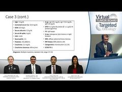
Ola Landgren, MD, PhD:Let’s go to the next case. I have another patient I would like to discuss. It’s a 64-year-old woman who presents with back pain. She has undergone imaging, and there’s evidence of a vertebral fracture in the fifth lumbar spine. This lady was previously completely healthy. Blood work is done showing a hemoglobin level of 12.3 g/dL. Her creatinine [level] is slightly elevatedat 2.0 mg/dL—so the creatinine clearance is estimated to be about 35 mL/minute. The LDH [lactic acid dehydrogenase level] is above the upper limit of normal, so increased beta-2-microglobulin is 7.1 mg/L, albumin is 3.4 g/dL, and the corrected calcium is determined to be 11.8 mg/dL. SPEP [serum protein electrophoresis] has been done and is negative. UPEP [urine protein electrophoresis] shows 7400 in 24-hour urine, and serum immunofixation picks up kappa light chains. The serum-free light-chain assay more accurately determines the concentrations of these light chains to be 2000 mg/L for kappa, and the lambda is estimated to be 20 mg/L.
Additional work-up is done. The first work-up was a skeletal survey. Additional work-up is done with a PET/CT [positron emission tomography or computed tomography], which shows a pathologic fracture in the L2. A bone marrow is done, showing 45% plasma cells. The bone marrow work-up also shows high-risk cytogenetics, including 17p deletions in more than 50% of the plasma cells. There’s also evidence of t(4;14), determined in more than 30% of cells. The clinical assessment is that this patient has an ECOG score of 1. Obviously the patient meets the definition for a diagnosis of multiple myeloma and is high-risk by the cytogenetic and FISH [fluorescence in situ hybridization] probes. By the revised International Staging System, this would be determined to be a stage III case.
Transcript edited for clarity.
Advertisement
Related Content
Advertisement


Advertisement
Advertisement
Trending on Targeted Oncology - Immunotherapy, Biomarkers, and Cancer Pathways
1
ASCO 2026 GU Cancer Highlights: Beyond the LBAs
2
Dr Tolaney on SG + Pembrolizumab Biomarker Data in First-Line mTNBC
3
FDA Approves Generic Eribulin Mesylate for Metastatic Breast Cancer
4
Bispecific ADC Iza-Bren Extends Survival in Advanced TNBC
5