Now Playing
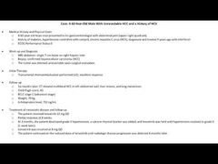
Ghassan K. Abou-Alfa, MD:Today, we’ll talk about a 60-year-old Asian man who presented to his gastroenterologist complaining of abdominal pain in the upper right upper quadrant. [He has] a history of diabetes and hypertension, which is controlled with Ramipril. Of note. he has chronic hepatitis C, which was diagnosed almost 9 years ago. He was treated initially with interferon, and thankfully his performance status is still goodECOG [Eastern Cooperative Oncology Group] 0.
An MRI [magnetic resonance imaging] of the abdomen took place and it showed a single 7-cm lesion in the right hepatic lobe. The biopsy confirmed the presence of hepatocellular carcinoma. Of note, surgery was not recommended, and was not the appropriate therapy because the tumor was deemed unresectable, mainly because of closeness to vascular invasion. And as such, the patients with locally advanced disease went ahead and received chemoembolization three times and with excellent response.
However, 6 months later a CT [computed tomography] scan showed that the liver tumors are worse. If anything, new liver lesions, abdominal wall extensive metastases, and lung metastases. Liver function was remained still good. Child-Pugh Score A. And if anything, the alpha-fetoprotein start elevating itself a little bit, 752, while at baseline it was close to about 250.
So the patient with the presence of metastatic HCC, BCLC [Barcelona-Clinic Liver Cancer staging system] or stage IV disease, went ahead and was started on lenvatinib, 12 mg daily. And after 8 weeks, there was clear evidence of partial response, and maybe about 9 months into the treatment patient started developing grade 3 hypertension. A calcium channel blocker was added, and the lenvatinib was held until the hypertension increase of the blood pressure improved and patient was restarted on the lenvatinib with a lower dose of 8 mg daily. And the patient continued on the reduced dose and really stayed on the therapy for another 6 months until progression of disease was noted.
So one might wonder, what do we learn from that study and what’s like so special about this case per se? I would like to start by talking about the, why did this patient get HCC to begin with? As we know, hepatitis C is what we always call “the silent epidemic.” Thankfully, nowadays there [are] a lot of therapies that can really treat the hepatitis C and actually cure from hepatitis C, but as we know this patient received interferon, which is not a curative intent, and if anything, patients might develop hepatocellular carcinoma if they had a chronic hepatitis, which is not cured, per se.
And it’s very important if a patient is aware of the presence of hepatitis C is to really seek medical attention and care, attempt hopefully at treating the hepatitis C, and more importantly, if they have a chronic hepatitis C, make sure they continue with their original screening to ensure that if there was any liver cancer to catch it rather earlier than later.
Understandably, we like to catch the disease in the early phases because curative intent, like for example, surgery, transplant, RFA [radiofrequency ablation] might be applicable, while, of course, like in this situation of our patient today, sadly, with the extent of disease [that] limits us from providing or offering any curative therapy.
Transcript edited for clarity.
A 60-Year-Old Male With Unresectable HCC and a History of HCV