Now Playing
EXPERT PERSPECTIVE VIRTUAL TUMOR BOARDBenjamin P. Levy, MD:Thank you for joining us for thisTargeted Oncology Virtual Tumor Board®, which is focused on advanced adenocarcinoma of the lung. In today’sTargeted Oncology Virtual Tumor Board®presentation, my colleagues and I will review 4 clinical cases. We will discuss an individualized approach to treatment for each patient and will review key trial data that impacts our decisions.
I’m Dr Benjamin Levy, an assistant professor of oncology and clinical director of medical oncology at the Johns Hopkins Sidney Kimmel Cancer Center, Sibley Memorial Hospital, in Washington, DC.
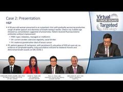
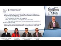
Today, I’m joined by Dr Gregory Riedlinger, an assistant professor of pathology in the Division of Translational Pathology at Rutgers Cancer Institute of New Jersey, of Robert Wood Johnson Medical School, Rutgers University, in New Brunswick, New Jersey; Dr Paul Paik, the clinical director of the Thoracic Oncology Service and assistant attending at Memorial Sloan Kettering Cancer Center in New York, New York; and Dr Anne Tsao, a director of both the mesothelioma program and thoracic chemoradiation program, and a professor in the Department of Thoracic Head and Neck Medical Oncology at the University of Texas MD Anderson Cancer Center, in Houston, Texas. Thank you for joining us. Let’s get started with our first case.
So, we’ll start our first case with a patient with locally advanced squamous cell nonsmall cell lung cancer. This is a 69-year-old male that was referred to pulmonology for symptoms of dyspnea and productive cough lasting for 4 months. In addition, he complained of mild dysphagia. He is a current smoker. He has a 40 pack/year history. His family history is consistent with a mother who died of cardiovascular disease and a father who had emphysema. The past medical history is consistent for hypertension and systemic lupus erythema, SLE or lupus, that’s been well controlled with no medications. The presentation with physical examthe patient showed pallor and decreased breath sounds on auscultation. His blood pressure was 100/60. All of his laboratory findings were within normal limits.
The patient, due to his symptoms, has a CAT scan. The scan reveals a 4-cm mass in the right upper lobe that extends into the right main bronchus and invades the mediastinum, and enlargement of the bilateral hilar and subcarinal lymph nodes. The patient undergoes a bronchoscopy with an EBUS [endobronchial ultrasound]-guided biopsy of the pulmonary mass and hilar lymph node, and the pathology is consistent with a grade 2 squamous cell carcinoma. His stage is IIIB. The patient has a brain MRI that is negative, and he goes on to receive a surgical consult. The surgeon deems the patient to be unresectable. The patient’s ECOG [Eastern Cooperative Oncology Group] performance status is 1. Due to unresectability, the patient is started on concurrent chemoradiation. He receives cisplatin/etoposide as the chemosensitization backbone with thoracic radiotherapy and achieves a partial response.
Transcript edited for clarity.