
|Articles|January 10, 2015
- Melanoma (January 2015)
- Volume 5
- Issue 1
Nanotechnology and Photodynamic Therapy Studied for Treatment of Metastatic Melanoma
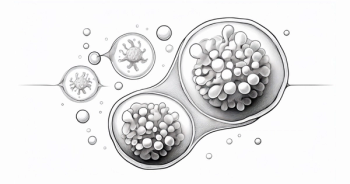
The combination of nanotechnology and photodynamic therapy may be useful in the future treatment of metastatic melanoma, according to researchers in the United States and Brazil.
Advertisement
The combination of nanotechnology and photodynamic therapy may be useful in the future treatment of metastatic melanoma, according to researchers in the United States and Brazil.
The increasing incidence of melanoma around the world has led to an estimated 230,000 new cases of melanoma worldwide since 2012. If diagnosed early, melanoma is treatable. However, as the disease progresses, the potential for cure becomes more remote. In addition, although approximately 91% of melanoma lesions occur on the skin, melanoma lesions on the eyes routinely account for 5% of cases, and those on mucosal surfaces account for approximately 1% of cases.1
If identified early, resected, and treated with a chemotherapeutic agents, such as dacarbazine, in a patient with good performance status, melanoma cure rates range from 97.9% to 99.8%. However, in later stages, the prognosis is substantially worse. The median survival time in patients with unresectable melanoma is approximately 28 months.1
Although newer targeted therapies are beginning to improve the prognosis of metastatic melanoma, further developments are needed. Victoria Monge-Fuentes, MD, of the department of genetics and morphology, University of BrasiÌlia, Brazil, and colleagues are investigating another way to target lesions: through a combination of drugs delivered using nanotechnology and activated by photodynamic therapy. One advantage of photodynamic therapy is its ability to stimulate certain excitation states of melanoma lesions. Through stimulation of oxidative damage, photodynamic therapy may exert anticancer effects, potentially stimulating direct killing of cancer tissue.1
Nanoparticles have been tested as both a drug delivery vehicle and as a method of targeting melanoma cells in murine models. Treatments that have been tested include nanoparticles that deliver medication preferentially to the site of tumors, nanoparticles that inhibit expression of genes in tumors through small interference RNA (siRNA) technology, and radiation-activated particles.2
Nanoparticles laden with chemotherapeutic agents have been tested in mouse models of melanoma. Investigators conjugated doxorubicin to fullerene nanoparticles and compared the efficacy of the nanoparticle formulation with the efficacy of free doxorubicin in a murine model of melanoma. The use of carbon nanostructures allowed scientists to load more than one-third of additional doxorubicin on each nanoparticle than is enabled by other systems. Fullerenes localized in the lysosomes of melanoma cells preferentially within 2 hours of administration. Free doxorubicin, unlike nanoparticle-formulated doxorubicin, tended to reduce the mass of mice spleen and heart tissue. These data suggest that preferential distribution to tumors through nanoparticle delivery of doxorubicin may reduce the risk of systemic adverse events (AEs).2,3
In mice, delivery of siRNA through nanoparticles has been used to successfully reduce expression of ribonucleotide reductase and c-Myc, a regulator oncogene, in melanoma cells. Current siRNA treatments include a cyclodextrin nanoparticle system that was recently investigated in a successful phase Ia/Ib clinical trial.4-6
In 24 patients enrolled in the Ia/Ib trial between May 2008 and September 2010, investigators increased the dosage of siRNA-laden nanoparticles from 3 mg/m2to 30 mg/m2. Enrolled patients had several types of cancer, including melanoma (n = 5), gastrointestinal cancer (n = 8), prostate cancer (n = 2), and other types (n = 9). Common AEs included chills and fever. Common severe AEs included lymphopenia, which occurred in 3 patients, and fatigue, which occurred in 2 patients. Less common severe AEs, occurring in 1 patient each, included hypersensitivity, ischemic colitis, diarrhea, and fever. Overall, the treatment was well tolerated, and with these results, trials are expected to progress to phase II.6
Although nanoparticles can carry chemotherapeutic agents and siRNA, they can also magnify the effects of radiotherapy. For example, nanoparticles may be activated by electromagnetic waves or sound. In mice, scientists have tested intratumoral administration of porphyrin-coated magnetic iron or iron (II, III) oxide nanoparticles. After murine melanoma cells were loaded with a dose of the iron particles each day for 3 days, mice were irradiated with 3 short pulses of external alternating magnetic fields. The treatments reduced the size of tumors, demonstrating that the technology may have some potential.7
With the development of many targeted agents for melanoma, use of nanoparticles may further enhance delivery to tumor cells in the future. In the near future, however, nanotechnology and photodynamic therapy are unlikely to be approved for melanoma therapy, overshadowed by other promising developments in combination treatment with immune checkpoint inhibitors, tumor-infiltrating lymphocyte therapies, and other immunotherapies.
References
- Monge-Fuentes V, Muehlmann LA, de Azevedo RB. Perspectives on the application of nanotechnology in photodynamic therapy for the treatment of melanoma.Nano Rev.2014;5. doi:10.3402/nano.v5.24381.
- Bombelli FB, Webster CA, Moncrieff M, Sherwood V. The scope of nanoparticle therapies for future metastatic melanoma treatment.Lancet Oncol.2014;15(1):e22-e32.
- Chaudhuri P, Paraskar A, Soni S, Mashelkar RA, Sengupta S. Fullerenol-cytotoxic conjugates for cancer chemotherapy.ACS Nano.2009;3(9):2505-2514.
- Davis ME, Zuckerman JE, Choi CH, et al. Evidence of RNAi in humans from systemically administered siRNA via targeted nanoparticles.Nature.2010;464(7291):1067- 1070.
- Chen Y, Bathula SR, Yang Q, Huang L. Targeted nanoparticles deliver siRNA to melanoma.J Invest Dermatol.2010;130(12):2790-2798.
- Zuckerman JE, Gritli I, Tolcher A, et al. Correlating animal and human phase Ia/Ib clinical data with CA- LAA-01, a targeted, polymer-based nanoparticle containing siRNA.Proc Natl Acad Sci U S A.2014;111(31):11449- 11454.
- Balivada S, Rachakatla RS, Wang H, et al. A/C magnetic hyperthermia of melanoma mediated by iron(0)/iron oxide core/shell magnetic nanoparticles: a mouse study.BMC Cancer.2010;10:119.
Articles in this issue
Advertisement
Related Content
Advertisement


Advertisement
Advertisement
Trending on Targeted Oncology - Immunotherapy, Biomarkers, and Cancer Pathways
1
FDA Accepts NDA for Daraxonrasib in Metastatic Pancreatic Cancer
2
Advanced Renal Cell Carcinoma: Tailoring Second-Line Therapy After Prior Immunotherapy
3
FDA Approves Zidesamtinib for Previously Treated ROS1-Positive NSCLC
4
Teclistamab/Talquetamab Reduces Risk of Progression by 89% in R/R Multiple Myeloma
5