
|Articles|July 9, 2021
Targeted Therapies in Oncology
- July 1 2021
- Volume 10
- Issue 9
Plasma Cell-Free Methylomes Offer Noninvasive Tumor Monitoring Across Cancer Settings
Author(s)Hayley Virgil
Th of plasma cell-free methylomes offers investigators a method to monitor tumor responses to treatment noninvasively, according to findings presented during the AACR Annual Meeting 2021.
Advertisement
The use of plasma cell-free methylomes offers investigators a method to monitor tumor responses to treatment noninvasively, according to findings presented during the AACR Annual Meeting 2021 in May.1 The method uses cell-free methylated DNA immunoprecipitation sequencing (cfMeDIP-seq) to detect and classify several types of cancer early on, according to Daniel D. De Carvalho, PhD, who presented the data.
The strategy has yielded positive results and has been shown to perform well in challenging applications such as early-stage renal cell carcinoma (RCC) and intracranial tumors, even helping to differentiate between distinct intracranial tumor entities.2
De Carvalho, the Helen M. Cooke associate professor in the Department of Medical Biophysics at the University of Toronto and the Canada Research Chair at the Princess Margaret Cancer Centre in Canada, further discussed the data supporting the use of cfMeDIP-seq and its potential for other low tumor burden clinical applications, such as monitoring for response and minimal residual disease (MRD).
Plasma Methylomes in Cancer Detection
The rationale for using plasma methylomes for cancer detection and monitoring is rooted in the fact that the cancer DNA methylation profile significantly differs from the normal tissue methylation profile, according to De Carvalho.
In one study, investigators examined DNA methylation variation in epigenetic domains in colorectal cancer (CRC) through the use of whole-genome bisulfite sequencing. By doing this, they identified a model for cancer that involved loss of epigenetic stability of well-defined genomic domains that underlies amplified methylation variability in cancer and may contribute to tumor heterogeneity.
“If you look at CpG islands…and CpG shores outside of the CpG islands, you can see a global increase in DNA methylation in cancer compared with normal [tissue] at hundreds of thousands of CpG sites,” De Carvalho said. “This provides a very rich set of biomarkers that one can use to monitor tumor DNA.”
Examining DNA Methylation Patterns to Understand Tissue of Origin Another notable area of information is the tissue of origin, De Carvalho said. A set of 500 CpG sites were utilized to identify different cancer types, including bladder, brain, breast, and colorectal tissue. Investigators were able to classify and discriminate these tissues from one another based on the DNA methylation profie alone.
As cells die and DNA is released into circulation, it is possible to capture floating cell-free DNA (cfDNA) and profile the methylome, De Carvalho said. Information content in the DNA methylation patterns can potentially be utilized to differentiate between normal tissue and cancer tissue and to differentiate the tissue of origin. Although this is an interesting concept, De Carvalho noted that several technical issues are associated with this strategy.
The Issue With Liquid Biopsies
“[We are faced with] a major needle-in-thehaystack problem with cfDNA,” De Carvalho explained. “In the real world, what we see is that 1 cancer cell has billions of letters of DNA….Somewhere between 10 to 20 million cell-free fragments can be released by a single cell as [it dies]. If you are looking for 1 event—1 KRAS hotspot mutation, [for example]—that is just 1 fragment out of potentially 50 million fragments per cell.”
This represents a significant barrier faced with liquid biopsies, especially with regard to early detection of residual disease and monitoring in situations with low tumor burden, according to De Carvalho.
“The way for us to think about this and try to solve for this is to take advantage of the DNA methylation. We have thousands of CpG sites of differential methylation,” De Carvalho noted. “[That means we] have a lot of needles in this haystack to look for [when] looking for methylation events.”
Investigators simulated different circulating tumor DNA (ctDNA) abundance, starting from 10% down to 0.001%, so high tumor burden vs low tumor burden, respectively.3 Thousands of genome-wide markers are needed to solve the needle-in-the-haystack problem when ctDNA abundance is less than 0.1%.
Detection of ctDNA is a function of ctDNA abundance, sequencing depth, and number of features investigated. For every fold decrease in tumor DNA abundance, it is important to have the same fold increase in the number of regions that are being searched for to maintain the probability of detection, according to De Carvalho.
Another challenge faced is that the human genome is depleted of CpG sites by deamination, De Carvalho said. “If you are trying to sequence the whole genome, about half of the potential cfDNA fragments being released by the cancer cell have no CpG, and, therefore, no DNA methylation [information],” De Carvalho added.
Investigators set out to completely separate methylated DNA from unmethylated DNA using an approach with monoclonal antibodies.4 “When you do that, compared with the expected distribution, most of the fragments we sequenced have a high number of CpG sites,” De Carvalho noted. He emphasized that this strategy only pulls down the methylated spikings, not the unmethylated spikings.
Differentiating Cancer Types With cfMeDIP-seq Utilizing this strategy, it is possible to discriminate between different cancer types. For example, in an analysis that examined the quality control of cfMeDIP-seq to differentiate between healthy donors and patients with lung cancer, breast cancer, CRC, leukemia, RCC, or bladder cancer, disease types could be differentiated based on the plasma cfDNA methylation.3
“You can see that you can have very distinct separation between the different cancer types,” De Carvalho explained. “Even if you have cancer cell lines that have no tumor microenvironment, only the methylation of the cell lines, you can see that they start to separate, as well, based on the signatures we identify in the plasma for these patients.”
A classifier was built using the original 388 plasma samples, and investigators validated an independent cohort of 199 samples. The validation set included samples from patients with lung cancer (n = 55), acute myeloid leukemia (n = 35), and pancreatic ductal adenocarcinoma (PDAC; n = 47). Notably a significant separation between the patient samples and samples from the healthy donors was observed. Moreover, even when dividing samples from patients with early-stage vs late-stage PDAC, it is possible to maintain the ability to discriminate between this disease and a healthy control sample, De Carvalho noted.
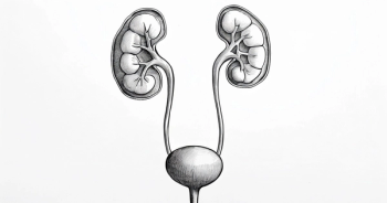
Investigators set out to further validate these findings within a cohort of patients with RCC.6 The trial featured 69 plasma samples collected from patients with RCC and 14 samples collected from healthy individuals who were cancer free.
“Using the plasma cell-free methylomes, there is very good separation between the cases and the controls in this cohort,” De Carvalho said.
“Even when compared with urothelial bladder cancer, you have a very good separation between both [this disease and RCC]. Interestingly, [we used] the same feature that we identify in the plasma if we profile with urine; we still have a very good separation between the cancer cases vs the control, just based on the cell-free DNA methylome profile of urine.”
Moreover, a cross validation was performed to further evaluate the ability to discriminate between cases and controls.
When using plasma cfDNA methylomes, investigators saw favorable classification discrimination between the RCC cases and the control samples, with a mean area under the receiver operating characteristic (AUROC) of 0.990 (95% CI, 0.985-0.995).
When looking at urothelial bladder cancer cases vs RCC cases in the plasma, very good discrimination was also observed (AUROC, 0.979; 95% CI, 0.956-0.991). Additionally, when examining discrimination between the RCC cases and the control in urine samples, investigators reported a mean AUROC of 0.858 (95% CI, 0.831-0.885).
Plasma cfDNA Methylomes Allow for Noninvasive Discrimination of Intracranial Tumors?
After establishing positive results in RCC, investigators moved on to examine the utility of cfMeDIPseq in a more difficult area: intracranial tumors. There is very little shedding of ctDNA in the plasma circulation with these tumors, according to De Carvalho. However, tissue-based DNA methylation profiling of these tumors can be utilized for molecular classification, De Carvalho explained.
“Many centers in Canada, Germany, and the United States have been using methylation profi ling from tumor DNA to classify the many different entities of intracranial tumors,” De Carvalho noted. “We tried to do the same, but with plasma cfDNA, because access to this tissue is highly invasive.”8
Investigators examined 447 plasma samples, 59 of which were glioma samples. A set of features, or differential methylated regions, helped discriminate these samples from other cancer types and healthy controls. These features were hypermethylated in the plasma of patients with glioma vs the plasma of the healthy controls, and they were also hypermethylated in the tumor of the patients with glioma vs the normal cfDNA.
A similar cross validation approach that was used in the RCC study was used in the glioma samples, according to De Carvalho. “Again, we had a very strong ability to discriminate between these [samples] vs other cancer types,” De Carvalho said.
Moreover, the ability to discriminate in the patient population was maintained in patients with IDH wild-type and IDH-mutant gliomas. These abnormalities do not result in a strong disruption in the methylation profile, and one can classify between both patient subgroups; this was also true with regard to low- vs high-grade gliomas.
“In increasing the number of samples from other intracranial tumor entities, such as meningioma [and others],” De Carvalho said, “you can see here that you have separation between them.”
Utilizing only the plasma cell-free methylomes, it was observed that the classifier does a fairly good job at identifying numerous intracranial tumor types, De Carvalho added.
Monitoring Response to Treatment Using ctDNA
In addition to helping identify and classify cancer types, cfMeDIP-seq may also have potential utility in cancer management, according to De Carvalho. In a population of patients with localized head and neck squamous cell carcinoma, investigators utilized an approach that incorporated the methylome profile, mutation profile, and fragment length.9
“In the patient samples compared with the healthy controls, the fragmental length of the methylated [ctDNA] starts to become [smaller] than the rest. If you look at the [patients with] healthy DNA vs the [patients with] mutant fragments…we can define the tumor based on mutation or methylation,” De Carvalho explained.
Mutations and methylation can be utilized independently to identify ctDNA, which indicates that the methylation signal allows one to quantify ctDNA as having a strong correlation between calculating the abundance based on mutation and percentage of ctDNA based on the signal of the methylome.
“Based on this, we can identify patients who are ctDNA positive vs negative and we can see a strong separation. Patients who are ctDNA negative have a much better overall survival [OS] than the [patients who are positive],” De Carvalho said.
Methylation also allows investigators to have independent prognostic markers in that methylation-specific events can be good or bad. Independent of that abundance of ctDNA, probability of OS can be calculated. Notably, patients with low methylation have a better survival than those who have a high methylation.
Lastly, cfMeDIP-seq can be monitored over time, as patients undergo treatment with different types of therapy. This can also help identify patients who have a clearance of ctDNA vs those who do not.
“Patients who are [clearing] up the ctDNA based on the methylation have a much better recurrence-free survival than the patients who do not [clear] their ctDNA. This is a very interesting application for MRD detection and monitoring of recurrence,” De Carvalho concluded.
REFERENCES
1. De Carvalho DD. Cancer early detection, classifi cation and monitoring using plasma cell-free methylomes. Presented at: AACR Annual Meeting 2021; May 17-21, 2021; virtual.
2. Hansen KD, Timp W, Corrada Bravo H, et al. Increased methylation variation in epigenetic domains across cancer types. Nat Genet. 2011;43(8):768-775. doi:10.1038/ng.865
3. Shen SY, Singhania R, Fehringer G, et al. Sensitive tumour detection and classifi cation using plasma cell-free DNA methylomes. Nature. 2018;563(7732):579-583. doi:10.1038/s41586-018-0703-0
4. Shen SY, Burgener JM, Bratman SV, De Carvalho DD. Preparation of cfMeDIP-seq libraries for methylome profi ling of plasma cell-free DNA. Nat Protoc. 2019;14(10):2749-2780. doi:10.1038/s41596-019-0202-2
5. Wilson ER, Helton N, Heath SE, et al. Genome-wide analysis of focal DNA hypermethylation in IDH-mutant AML samples. bioRxiv. Published online March 3, 2021. doi:10.1101/2021.03.03.433799
6. Nuzzo PV, Berchuck JE, Korthauer K, et al. Detection of renal cell carcinoma using plasma and urine cell-free DNA methylomes. Nat Med. 2020;26(7):1041-1043. doi:10.1038/s41591-020-0933-1
7. Capper D, Jones DTW, Sill M, et al. DNA methylation-based classifi cation of central nervous system tumours. Nature. 2018;555(7697):469474. doi:10.1038/nature26000
8. Nassiri F, Chakravarthy A, Feng S, et al. Detection and discrimination of intracranial tumors using plasma cell-free DNA methylomes. Nat Med. 2020;26(7):1044-1047. doi:10.1038/s41591-020-0932-2
9. Burgener JM, Zou J, Zhao Z, Shen SY, De Carvalho DD, Bratman SV. Comprehensive detection of ctDNA in localized head and neck cancer by genome- and methylome-based analysis. Clin Cancer Res. 2020;26(suppl 11):PR13. doi:10.1158/1557-3265.LiqBiop20-PR13
Articles in this issue
about 5 years ago
New Guidelines Divide Recommendations for Driver and Nondriver NSCLCabout 5 years ago
After PD-1 Inhibitor Indications Are Withdrawn in SCLC, What Now?about 5 years ago
Integrating Remote Symptom Management With ePROs in Cancer Careabout 5 years ago
Novel Approaches Address Tumor Heterogeneity in GI Malignanciesabout 5 years ago
Molecular Profiling Is the Future of Biliary Tract TreatmentAdvertisement
Related Content
Advertisement


Advertisement
Advertisement
Trending on Targeted Oncology - Immunotherapy, Biomarkers, and Cancer Pathways
1
Advanced Renal Cell Carcinoma: Summarizing a Practical First-Line Treatment Algorithm
2
Long-Term OMNIVORE Data Show Durable Treatment-Free Survival in RCC
3
On-Body Injector Technology Streamlines Subcutaneous Myeloma Therapy
4
First Patients Dosed With TLX597-Tx in Trial for mHSPC
5