|Videos|September 12, 2018
Case 2: Stage IV PD-L1-Negative, KRAS-Mutant Lung Adenocarcinoma
Advertisement
Episodes in this series


EXPERT PERSPECTIVE VIRTUAL TUMOR BOARDPaul K. Paik, MD:We’re going to switch gears now to our second case, which is a stage IV lung adenocarcinoma patient whose tumor is PD-L1 [programmed-cell death ligand 1]-negative and harbors aKRASmutation. This will help us talk about some of the newer data that’s come out over the past 6 months, that is really relevant in terms of the change in the first-line landscape for patients with stage IV non­small cell lung cancer.
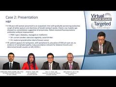
The patient is a 58-year-old woman. She presented to the outpatient clinic with progressive cough and shortness of breath, which happened over the past few weeks. The emergency [department] doctors had gotten a chest X-ray, which showed left lower-lobe consolidation. And so, the thought was that the patient had pneumonia. She was treated with fluoroquinolone antibiotics but there was no clinical improvement in her symptoms. On the examination, at the time, she was noted to have a past medical history of type 2 diabetes. She was a current smoker. She was on metformin. She exercised regularly, was a social drinker, and had a family historya maternal grandmother who had died of breast cancer.
On examination, at the time, she did actually look ill. She was tachypneic. The O2saturation did show some diminution. She was 93% on room air. On examination, there was no lymphadenopathy. There were bilateral rhonchi and diminished breath sounds on the left side, commensurate with what they found on the chest x-ray. Her ECOG [Eastern Cooperative Oncology Group] performance status was 1. Laboratory findings did show leukocytosis. The white cell count was 16, and the ABG [arterial blood gas] interpretation showed respiratory alkalosis along with a decrease in the partial pressure of the oxygen. Sputum blood and urine cultures were negative. This was followed with a CT scan of the chest. Here, we found a fairly large 8-cm left lung mass, consolidation with air bronchograms, some adjacent tree-in-bud nodules, no effusions, and the major airways were patent.
The patient subsequently got a bronchoscopy with washing. The washing showed atypical cells, but the transbronchial biopsy showed a high-grade lung adenocarcinoma that was largely micropapillary, in terms of histologic features. The testing that was done was sort of limited sequencing. This was not next-generation sequencing. An Oncomine panel did detect aKRAScodon 12 mutation, anEGFRmutation, and wild-typeBRAF. The patient was also wild-type forALKandROS1on FISH [fluorescence in situ hybridization] testing. The IHC [immunohistochemistry] did show TTF1, commensurate with the lung adenocarcinoma diagnosis. The PD-L1, in this case, was 0%. And finally, to round out the staging workup, the patient had a PET scan. In addition to the left lung mass, which was FDG-avid, there was also a right adrenal gland that was FDG-avid, clinching a diagnosis of stage IV lung adenocarcinoma.
Transcript edited for clarity.
Advertisement
Related Content
Advertisement


Advertisement
Advertisement
Trending on Targeted Oncology - Immunotherapy, Biomarkers, and Cancer Pathways
1
FDA Accepts NDA for Daraxonrasib in Metastatic Pancreatic Cancer
2
Allogeneic CD70 CAR T-Cell Therapy Shows Activity in Refractory Kidney Cancer
3
FDA Approves Zidesamtinib for Previously Treated ROS1-Positive NSCLC
4
Indefinite Myeloma Maintenance Shows No OS Benefit vs 2-Year Duration
5