Now Playing
Andrew G. Gianoukakis, MD: Hello, I’m Dr Andrew Gianoukakis, professor of medicine at the Harbor-UCLA Medical Center in Torrance, California. I’m here today to discuss a case of thyroid cancer with you and to go through the appropriate management, including the administration of systemic therapy as needed.
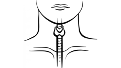
The case is a 73-year-old woman with differentiated thyroid cancer. The patient initially complained of a lump in her neck with occasional swelling and dysphagia. Her past medical history includes only obesity with a BMI [body mass index] of 32. Her physical examination is notable only for a palpable nontender neck mass.
Laboratory testing indicated a TSH [thyroid stimulating hormone] level of 1.1 [µU/mL]—within normal limits. All other testing was likewise, within normal limits. An ultrasound of the neck revealed a 3-cm mass in the right lobe of the thyroid. In addition, several suspicious lymph nodes were noted ranging from 0.2 to 0.8 cm in size. An ultrasound-guided FNA [fine needle aspiration] biopsy of the 3-cm right lobe mass was performed, and papillary thyroid carcinoma was diagnosed.
The patient subsequently underwent a total thyroidectomy with bilateral central neck dissection. The pathology revealed a 3-cm classic papillary thyroid cancer in the right lobe of the thyroid. Additionally, 2 of 5 central compartment lymph nodes removed were noted to be positive for thyroid cancer, the largest measuring 1.4 cm. The patient was staged as a T2N1MX. Her ECOG performance status was noted to be 0.
After her biopsy and surgery, the patient was treated with radioactive iodine. She received an ablative dose, and levothyroxine suppression was added to her treatment regimen. At 6 months, her thyroglobulin level was noted to be 4 ng/mL with a TSH level of 0.4 [μU/mL]. A neck ultrasound was performed and noted to be unremarkable. At 1 year, her TSH level remained suppressed at 0.3 [μU/mL], yet her thyroglobulin level had increased to 18 ng/mL. A follow-up ultrasound of the neck was unremarkable, and therefore a CT scan was performed. Ten lung nodules were noted with the largest being 1.2 cm in size. A second treatment of radioactive iodine was administered. The post-therapy scan did not disclose any lung or other iodine uptake.
At year 2, her thyroglobulin level had now risen to 30 [ng/mL] with a TSH level remaining suppressed at 0.3 [μU/mL]. Follow-up chest CT showed an increasing number and size of thyroid nodules; the largest thyroid nodule had increased to 1.6 cm.
The patient remains asymptomatic. Consideration of additional therapy is appropriate at this time, including the potential for systemic therapy.
The case is a pretty typical case of an older person presenting with a thyroid nodule diagnosed to be thyroid cancer. The patient’s disease appears to have progressed rather quickly to metastatic disease. The administration of a second dose of radioactive iodine, which disclosed no uptake in the macroscopic lung lesions, in addition to lung lesions at year 2 that continue to grow, render the patient radioiodine refractory. The patient was appropriately treated initially with surgery and an ablative dose of radioactive iodine and appropriately followed with neck ultrasound and thyroglobulin testing. I would only add that as part of the thyroglobulin testing, we typically test for thyroglobulin antibodies, which allow us to have confidence in the thyroglobulin measurement. The thyroglobulin antibodies—when positive, and they can be positive in up to about 25% of patients with thyroid cancer—can interfere with the thyroglobulin assay.
Transcript edited for clarity.
Case: A 73-Year-Old Woman With Differentiated Thyroid Cancer
Initial Presentation
Clinical Work-up and Initial Treatment
Subsequent Treatment and Follow-up