
|Articles|August 14, 2021
Targeted Therapies in Oncology
- August 2021
- Volume 10
- Issue 11
Nanotechnology’s Reach Enhances Drug Delivery
Author(s)Thao K. Adams, PharmD, BCOP
Nanotechnology advances in oncology have applications in diagnosis, biomarker screening, and most notably, treatment.
Advertisement
Nanotechnology is a rapidly evolving treatment modality in oncology.1 Nanotechnology advances in oncology have applications in diagnosis, biomarker screening, and most notably, treatment. Conventional chemotherapy has been the mainstay of oncology treatment; however, due to its indiscriminate mechanism of inducing cytotoxicity, multiple undesired toxicities are experienced.2 Compared with conventional chemotherapy, nanotechnology has the potential to be precise and effective in selecting for tumor cells with a reduction in undesired adverse events (AEs) on normal tissue, making this novel approach a promising treatment in cancer.1
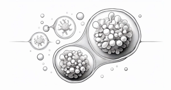
Nanoparticles are defined as particles with a size of 1 to 100 nm.3 The optimal diameter range to achieve enhanced permeability and retention (EPR) effects is 10 to 100 nm for best drug delivery.2 Nanoparticles can be composed of organic, inorganic, and hybrid material.
Organic nanoparticles include liposome and polymer-based compositions. Inorganic nanoparticles include gold, carbon nanotubes, silica, magnetic, and quantum dots. Inorganic nanoparticles have higher surface area to volume ratio with an easily modifiable surface conjugation chemistry; however, its biocompatibility and biodegradability is not as robust as organic nanoparticles. Within inorganic nanoparticles, silica nanoparticles are favorable carriers for drug delivery due to its pharmacokinetics, treatment efficacy, and high stability.
Hybrid nanoparticle composition may include lipid-polymer, organic-inorganic, and cell membrane-coated.1,2,4 In drug delivery, passive targeting of tumor cells using nanotechnology relies on the size of the nanoparticle to be retained by cancer cells with poor lymphatic drainage. Alternatively, using ligands on the surface of nanoparticles to directly target tumor cells is a modality in active targeting.2
“Nanotechnology is a rather big field and it is a broad field. There are many people working on trying to focus on using nanoparticles for clinical application in oncology,” Maciej Lesniak, MD, at the Robert H. Lurie Comprehensive Cancer Center of Northwestern University, said during an interview with Targeted Therapies in Oncology.
Lesniak, the Michael J Marchese Professor and Chairman, Department of Neurological Surgery, at Northwestern University Feinberg School of Medicine in Chicago, and program leader in Neuro-Oncology at Northwestern, was an investigator evaluating the use of spherical nucleic acids (SNAs). The SNA is a type of oligonucleotide, such as small interfering RNA (siRNA), which surrounds a nanoparticle core. The nanoparticle core can be made from gold, silver, iron oxide, quantum dots, platinum, silica, core-shell, and liposomes.2 In this particular study, SNAs containing siRNA oligonucleotides on gold nanoparticle cores were used to target BCL2L12, a protein coding gene, for the treatment of recurrent glioblastoma.4 There were 8 patients enrolled with the study agent, NU-0129, administered intravenously 24 to 28 hours prior to tumor resection and tissue analysis. Grade 3 or higher AEs potentially related to administration of NU-0129 were hypophosphatemia and lymphopenia, the investigators reported. Only 6 of 8 tumor resections were analyzed due to tissue sample size.4
“We were able to show that peripheral injection of the gold nanoparticles actually achieves distribution within the brain and the tumor itself and that it seems to downregulate [one of the key] proteins within the tumor itself,” Lesniak said. Results demonstrated gold nanoparticle accumulation in tumor-associated endothelium in tumor- associated macrophages and Ki67-positive tumor cells.4 Downregulation of BCL2L12 protein signified drug penetration of the blood brain barrier and accumulation in tumor cells. Acute and long-term toxicities of NU-0129 were not experienced in patients.4
Gold Nanoparticles Gold nanoparticles are an attractive treatment option because it can be used for gene therapy, photothermal therapy, and immunotherapy.2 Gold nanoparticles can be used to decrease the time of disease metastasis by increasing nuclear membrane stiffness. It may be modified to contain methoxy-polyethylene glycol thiol (PEG), RDG peptides, and nuclear localization signal peptides. The tumor cells absorb the gold nanoparticles and subsequently cause nuclear stiffness of the cancer cell to decrease the rate of metastasis of cancer cells.3
In addition to gold nanoparticles, other metals have been used in combination with magnetic fields.2 Magnetic nanomaterials is a therapeutic option that has demonstrated promising results.6 The advantage associated with magnetic nanomaterials is the destruction of cancer cells via hyperthermia and mechanical force, providing an alternative to chemotherapy. Additionally, a magnetic field may be used to initiate specific signaling pathways to cause tumor cell lysis without contact. In a study evaluating the effects of magnetic nanoparticles on glioma in mice implanted with U87 glioma cells. Treatment involving rotating magnetic fields were provided daily to the treatment group.6
“We looked at using magnetic nanoparticles that oscillate upon application of external fields. When you bind these nanoparticles to cancer cells, just the gentle oscillation disrupts the cancer cell membrane … and therefore mechanical destruction of cancer cells,” Lesniak, an investigator in the trial, said.
The treatment group achieved tumor regression by day 7 whereas the control group had tumor growth.6 Additionally, by day 28, 40% of the treatment group demonstrated effective treatment strategy with effective tumor growth inhibition. Long-term tumor growth suppression was also observed in 40% the treatment group with long-term survival benefit. The results from this study provided a different modality in treating glioma with mechanical force. Another advantage demonstrated from the study with the use of magnetic field is the level of penetration to the tumor cells without damaging other biological tissues.6
On the advantages of nanotechnology, Lesniak said, “the advantages are obvious. There is an intellectual, intrigue component to these nanoparticles [and their potential use]. There’s an element of excitement about nanoparticles and its applicability in the form of cancer.”
The versatility of nanoparticles allows for its application with multiple oncology treatments including, chemotherapy, targeted therapy, radiotherapy, hyperthermia, gene therapy, and immunotherapy.2 Due to the properties of nanoparticles, self-assembly, stability, and specificity are advantages of its use, allowing for interactions with tumor cells and the tumor microenvironment with improved specificity compared with chemotherapy.2,4
Additional Benefits
Other benefits thought to derive from nanotechnology is the ability to enhance immunotherapy and reverse the tumor immunosuppressive microenvironment.2 Furthermore, nanotechnology may be used to overcome multidrug resistance, especially with hybrid nanoparticles using multiple chemotherapy agents within the carrier. There have been reports of nanoparticles overcoming multidrug resistant tumors in breast cancer, ovarian cancer, and prostate cancer.2
Alternatively, Lesniak said, “the biggest challenges [with nanotechnology] have to do with the manufacturing and preclinical development. For instance, gold [nanoparticles] work great in some preclinical models and in vitro. However, it’s somewhat difficult to execute in human trials simply because of the cost associated with producing gold nanoparticles. Most [investigators] start switching to lipid nanoparticles or other chemical formulations that would allow scalability and would therefore decrease the cost associated with the production that would be necessary to enroll a large number of patients.”
Lesniak also commented that another notable challenge with nanoparticles is the effects on the rest of the organ systems. Nano-bio interactions is complex with preclinical data demonstrating only 0.7% of injected nanoparticles accumulating in tumors.7 “We need more human clinical trial work, especially in cancer, to [understand when nanoparticles are cleared from the body], how they are cleared, and the [clinical implications]. We always want to balance therapy with preventing harm to organs and this is a challenge. It is an area of active investigation,” Lesniak said.
Due to the practicality, the majority of nanoparticles is in the form of liposomes.7 Liposomes are spherical nanoparticles with an outer lipid layer mimicking human cells enclosed around an aqueous center, which may contain chemotherapy and nucleic acids.1,2,4 One of the successful clinical applications of liposomes is with liposomal doxorubicin.4 Due to the nanoparticle system, liposomal doxorubicin has demonstrated less cardiotoxicity compared with conventional doxorubicin. Likewise, nanoparticle albumin-bound paclitaxel also exhibits less AEs compared with conventional paclitaxel, achieving the goal of decreased toxicity with nanoparticle use.2
Drug Design Lesniak commented on how to incorporate nanotechnology into drug design. “These translational studies that go from bench to bedside where we develop a clinical grade agent, work with FDA to get [approval and an infrastructure that is compliant with regulatory issues] and test it in early stage clinical trials patients, whether it’s gene therapy or nanoparticle protocols, involves a team approach in working with a disease for which there is an unmet need.”
Current and future work in nanotechnology may include “looking at the immunomodulatory function and how to use nanoparticles to enhance the immune system to fight cancer,” said Lesniak. Furthermore, he stated, “one area where I see [nanotechnology] potentially making a big dent is in terms of imaging micrometastases. Thinking about how we can detect smaller tumor burdens in the order of one thousand cells instead of a mass of millions of cells is a really important thing when it comes to metastases. We can then treat those [micrometastases] with targeted radiotherapy or chemotherapy [using nanotechnology].”
Although nanotechnology in the treatment of cancer has tremendous potential, the clinical implications have yet to be fully manifested. Overcoming the nano-bio barriers in a team approach to utilize advanced disease modeling, omics technologies, computing, and using artificial intelligence may accelerate the research and translational medicine into successful clinical settings.7 “We still have a ways to go and I think the incredible people around the world, including this country, will pave the way for the future,” Lesniak concluded.
References:
1. Jin C, Wang K, Oppong-Gyebi A, Hu J. Application of nanotechnology in cancer diagnosis and therapy - a mini-review. Int J Med Sci. 2020;17(18):2964-2973. doi:10.7150/ijms.49801
2. Yao Y, Zhou Y, Liu L, et al. Nanoparticle-based drug delivery in cancer therapy and its role in overcoming drug resistance. Front Mol Biosci. 2020;7:193. doi:10.3389/fmolb.2020.00193
3. Leng F, Liu F, Yang Y, Wu Y, Tian W. Strategies on nanodiagnostics and nanotherapies of the three common cancers. Nanomaterials (Basel). 2018;8(4):202. doi:10.3390/nano8040202
4. Wang X, Yang L, Chen ZG, Shin DM. Application of nanotechnology in cancer therapy and imaging. CA Cancer J Clin. 2008;58(2):97-110. doi:10.3322/CA.2007.0003 5. Kumthekar P, Ko CH, Paunesku T, et al. A first-in-human phase 0 clinical study of RNA interference-based spherical nucleic acids in patients with recurrent glioblastoma. Sci Transl Med. 2021;13(584):eabb3945. doi:10.1126/scitranslmed.abb3945
6. Cheng Y, Muroski ME, Petit DCMC, et al. Rotating magnetic field induced oscillation of magnetic particles for in vivo mechanical destruction of malignant glioma. J Control Release. 2016;223:75-84. doi:10.1016/j.jconrel.2015.12.028
7. de Lázaro I, Mooney DJ. Obstacles and opportunities in a forward vision for cancer nanomedicine. Nat Mater. 2021;10.1038/s41563021-01047-7. doi:10.1038/s41563-021-01047-7
Articles in this issue
Advertisement
Related Content
Advertisement


Advertisement
Advertisement
Trending on Targeted Oncology - Immunotherapy, Biomarkers, and Cancer Pathways
1
CMS Proposes Cut to 340B Drug Payments in 2027 Outpatient Rule
2
FDA OKs ProstACT Trial Part 2 Protocol for Metastatic Prostate Cancer
3
The Growing Rationale for Earlier Targeting of GPRC5D in Multiple Myeloma
4
Botensilimab Plus Balstilimab Yields 33% OS Rate at 3 years in MSS mCRC
5