|Articles|November 28, 2019
Understanding Virus-Induced Cancers Can Elucidate Precancerous Lesion Development
Author(s)Erin M. Burns, PhD, MSPH
Approximately 20% of cancers worldwide are linked to an infectious agent. Currently, there are seven known oncogenic viruses, which include Epstein-Barr virus, hepatitis virus B and C, human papillomavirus, human T cell lymphoma virus 1, Kaposi sarcoma virus and Merkel cell polyomavirus. Among these agents, HBV, HCV and HPV each contribute to ap- proximately 5% of all cancer cases.
Advertisement
Robert L. Ferris, MD, PhD
Approximately 20% of cancers worldwide are linked to an infectious agent. Currently, there are seven known oncogenic viruses, which include Epstein-Barr virus (EBV), hepatitis virus B and C (HBV and HCV), human papillomavirus (HPV), human T cell lymphoma virus 1 (HTLV-1), Kaposi sarcoma virus (also known as human herpes virus 8; KSVH or HHV8) and Merkel cell polyomavirus (MCPyV ). Among these agents, HBV, HCV and HPV each contribute to approximately 5% of all cancer cases.1,2
HBV and HCV contribute to liver cancer development, and HCV can also cause non-Hodgkin lymphoma. There are at least 12 strains of HPV that can contribute to the development of anal, cervical, penile, throat, vaginal and vulvar cancer. EBV increases the risk for developing Burkitt lymphoma, Hodgkin and non-Hodgkin lymphoma, and stomach cancer. HTLV-1 has been linked with the development of adult T cell leukemia/lymphoma. HHV8 is correlated with Kaposi sarcoma in immunocompromised individuals, and MCPyV has been associated with the development of Merkel cell carcinoma.
EBV, HPV, HTLV-1, KSVH and MCPyV are classified as direct carcinogenic pathogens; a critical portion of the viral genome can be detected in each cancer cell, which leads to viral oncogene expression that disrupts cell-cycle checkpoints, inhibits apoptosis and facilitates cell immortalization. Indirect carcinogenic pathogens, including HBV and HCV, are not associated with the induction of oncogene expression. Instead, persistent infection contributes to a chronic inflammatory state that leads to the release of cytokines, chemokines, prostaglandins and reactive oxygen species, which can result in immune system dysregulation and the promotion of neovascularization.
Despite contributing to the global cancer burden, oncogenesis is an uncommon outcome of infection and is not in line with the general life cycle of these oncogenic pathogens. When it occurs, pathogen-induced oncogenesis may be observed years after the initial infection. This long time to onset demonstrates the requirement for additional steps other than infection by the pathogen alone.3,4
Increased rates of pathogen-driven cancers have been reported in areas with increased infection rates, including underserved communities and developing countries, and in immunosuppressed populations. In a meta-analysis of patients with HIV/AIDS and transplant recipients, there was a significantly increased incidence of cancer, including EBV-related lymphoma/leukemia, HBV- and HCV-associated hepatocellular carcinoma and HPV-positive cervical cancer.5However, incidence rates of most common epithelial cancers were less than or equal to that observed in the general population. These data suggest that immunodeficiency may contribute to the observed increased cancer incidence in these populations.
“The fundamental premise of cancer immunotherapy is that the immune system fails to identify premalignant lesions that can progress into cancer,” explainedRobert L. Ferris, MD, PhD, director of UPMC Hillman Cancer Center, Hillman professor of oncology, associate vice chancellor for cancer research, and co-director of the Tumor Microenvironment Center in Pittsburgh. “Viruses provide an ideal model, in a way, to understand the failure of the host immune system to control a precancerous lesion, in this case, driven by oncogenic viruses.” Ferris co-chaired the concurrent session on virus-driven cancers during the Society for Immunotherapy of Cancer’s 34th Annual Meeting (SITC 2019).
According to Ferris, commonalities need to be drawn regarding the similarities and differences of viruses, the identification of viral gene products capable of transforming normal cells into malignant cells and the contributions of viruses to metastasis and invasion.
“As we’ve come to learn, one of the key features of viruses that can cause cancer is the ability to downregulate or escape the normal immune system response, permitting them to grow unchecked while they turn their target cell into a precancerous lesion,” said Ferris. “We have tried to identify ways to incorporate novel, innovative diagnostic or therapeutic strategies and combinations to reactivateand I think that’s one of the keys—the antiviral immunity as a mechanism of treatment.”
During his training, Ferris was involved in work on cancer vaccines against HPV, working at Johns Hopkins University where investigators initially made the observation that HPV was present in head and neck cancers. “That was sort of what got me very interested in immunotherapy of head and neck cancers because you had a target; something to shoot at,” explained Ferris.
In a two-year update of long-term survival of patients treated with nivolumab (Opdivo) or investigator’s choice for recurrent or metastatic squamous cell carcinoma of the head and neck, the 24-month overall survival rate was nearly three times higher with nivolumab therapy than it was with investigator’s choice, regardless of tumor HPV status. Studies are ongoing to investigate the combination of HPV-targeted immunotherapy and anti-PD-1 in head and neck cancer.6
Traditionally, head and neck cancer development is associated with excessive tobacco and alcohol use.7,8Patients with HPV-positive cancers tend to have better outcomes. “It’s unfortunate that these folks contract these cancers, but I should point out that HPV in head and neck cancer has a dramatically better prognosis than HPV-negative cancer,” explained Ferris. Areas of future investigation include determining why HPV-positive head and neck cancer patients have approximately twice the survival of patients with HPV-negative tumors.
It is important to remember that not all virus-induced cancers arise or are at an increased frequency in situations of immunosuppression. However, this is the case for Merkel cell carcinomas. The factors that contribute to Merkel cell carcinoma development are not fully understood, but the incidence of Merkel cell carcinoma is higher in immunosuppressed and elderly individuals than it is in immunocompetent and younger individuals.
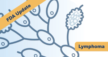
Merkel cell carcinoma develops when Merkel cells, which are found in the epidermis near nerve endings, grow out of control (FIGURE). It is a rare neuroendocrine tumor that had an estimated incidence rate of 0.18 to 0.41 per 100,000 population in 2006. However, over the last 20 years the incidence rate of Merkel cell carcinoma has increased to approximately 0.7 cases per 100,000 person-years in the United States in 2013. This increase has been attributed to improved diagnostic tools and an increasingly aged population based on the exponentially increasing incidence of Merkel cell carcinoma with age.9-11, 12-14
Risk factors include high levels of natural and artificial sunlight exposure, a history of other cancer diagnoses, age greater than 50 years, male sex, white race and having a weakened immune system due to disease or immunosuppressive drugs (such as after organ transplantation).9
Approximately 10% of patients with Merkel cell carcinoma have chronic immunosuppression, which is a greater rate than that seen in the general population. Furthermore, patients with immuno-suppressed Merkel cell carcinoma have significantly reduced cancer-specific survival rates than patients with non-immunosuppressed Merkel cell carcinoma (40% versus 74% at three years, respectively). These data suggest that immuno-suppressed patients have an increased likelihood of developing Merkel cell carcinoma and they are more likely to die from the disease, which high- lights the importance of immune regulation in this virus-driven cancer.15
“Until very recently, we did not have any effective therapies that could prolong life for people whose Merkel cell cancer had spread to the point where it could not be removed surgically,” explained Suzanne L. Topalian, MD, Bloomberg-Kimmel professor of cancer immunotherapy, professor of surgery and oncology at Johns Hopkins University School of Medicine, director of the melanoma program at the Kimmel Cancer Center and associate director of the Bloomberg-Kimmel Institute for Cancer Immunotherapy, all in Baltimore, Md.
“Those patients were typically treated with combination chemotherapy or radiation therapy, but their prognosis was still very limited, with life expectancy, on average, less than a year after the diagnosis of advanced metastatic Merkel cell carcinoma,” said Topalian, who presented during this concurrent session. “A real revolution in how we treat Merkel cell cancer came when antiPD-1 and PD-L1 drugs were tested against this disease.”
In 2016, in a phase 2 study of 26 patients with advanced Merkel cell carcinoma who had not received previous systemic therapy and were treated with pembrolizumab (Keytruda), the objective response rate (ORR) was 56% among 25 patients with one or more evaluations.16 Among these patients, four had a complete response and 10 had a partial response. Over a median follow-up of 33 weeks, two of the 14 patients who had a response experienced relapse, with a response duration ranging from 2.2 months to 9.7 months. The six-month progression-free survival rate was 67%. Among the 16 patients with MCPyV-positive tumors, the response rate was 62%, whereas it was 44% among those with virus-negative tumors.
In a multicenter, open-label, phase 2 study, 88 patients with stage IV chemotherapy-refractory, histologically confirmed Merkel cell carcinoma were treated with at least one dose of avelumab (Bavencio)).17After a median follow-up of 10.4 months, 32% of patients achieved an objective response, including eight complete responses and 20 partial responses. At the time of analysis, 82% of responses were ongoing (TABLE).17
“Both of those trials showed substantial response rates to these drugs, and they indicated in early results that the responses appeared to be durable,” noted Topalian. These studies have recently reported long-term follow-up data, confirming the durability of many responses.
Of 50 patients treated with pembrolizumab for up to two years, the ORR was 56%, with an ORR of 59% in virus-positive tumors and an ORR of 53% in virus-negative tumors. The 24-month progression-free and overall survival rates were 48.3% and 68.7%, respectively. Treatment-related adverse events of grade 3 or higher were reported in 28% of patients, leading to discontinuation of treatment in 14% of patients, with one patient death.18
Among 88 patients treated with avelumab and followed for at least 12 months, there was a confirmed ORR of 33%, including 11% complete responses. Approximately 74% of responses lasted at least one year, with 72.4% of responses ongoing at the time of analysis. The median duration of response was not reached. The one-year progression-free and overall survival rates were 30% and 52%, respectively. Durable responses were reported irrespective of MCPyV status.19
“This is really great news for patients with this disease, but what we learned scientifically, and what we published in 2016, was that responses to antiPD-1 treatment were similar in the two major groups of Merkel cell cancers, which are those cancers that are caused by the Merkel cell polyomavirus and those that are not,” explained Topalian.
A study designed to evaluate the genetic basis of Merkel cell carcinoma via exome sequencing reported that MCPyV-negative tumors had a high mutation burden, with a median of 1,121 somatic single nucleotide variants (SSNVs) per tumor, whereas MCPyV-positive tumors had a low mutation burden, with a median of 12.5 SSNVs per tumor. Additionally, MCPyV-negative Merkel cell carcinomas harbored more predicted tumor neoantigens than melanomas or non-small cell lung cancers, for which immune checkpoint blockade has been shown to lead to durable clinical responses. These data suggest that these tumors would be good targets for immunotherapy because the high expression of neoantigens should lead to stronger immune responses and tumor rejection.20
“I would say that Merkel cell cancers that are virus-positive really break that mold because they don’t have lot of mutations,” said Topalian. “But what they do have are viral proteins, which are very strong immunogens. This cancer teaches us that it’s not only the number of tumor antigens that is important for cancer rejection, it’s the quality of those antigens, and how strong they are, in terms of how highly they provoke an immune response.”
Current studies in Merkel cell carcinoma are focused on improving the results that have been reported with antiPD-1 and anti–PD-L1 therapy. Biomarkers are needed to help identify the patients that are most likely to respond or not respond to therapy. Additionally, studies are needed to elucidate the mechanisms contributing to treatment resistance in order to develop more powerful combination therapies.
Additional studies are underway using immunotherapy to treat patients with Merkel cell carcinoma at earlier stages with adjuvant or neoadjuvant therapy. “We reported some preliminary results with the antiPD-1 drug, nivolumab, in the neoadjuvant setting at ASCO [American Society of Clinical Oncology] 2018,” explained Topalian. “We saw a high rate of pathologic responses in the resected tumor specimens and we found that result very promising, so now there are other trials ongoing to look at neoadjuvant forms of immunotherapy in Merkel cell cancer.”
PD-L1 and microsatellite instability (MSI), which is associated with a very high tumor mutational burden, are two biomarkers that are FDA-approved for directing antiPD-1 or anti– PD-L1 therapy in certain cancer types. However, in Merkel cell carcinoma, a high mutation burden will not inform selection of patients for therapy because there are high response rates in virus-positive tumors, which have a low tumor mutational burden, and in virus-negative tumors, which have a high tumor mutational burden.
“We have also published that the PD-L1 marker doesn’t seem to help us here,” explained Topalian. “In our studies of pembrolizumab treatment in the advanced disease setting, PD-L1 expression in pre-treatment tumor biopsies did not correlate with response, so that really means that we need to look at some different markers for Merkel cell cancer. That is a very active area of research.”
According to Topalian, three things to note about Merkel cell carcinoma and virus-induced cancers are that viral antigens can act as tumor rejection antigens, a high tumor mutational burden is not required for response to antiPD-1 therapies, and applying immunotherapy in earlier disease settings may be advantageous.
The Future of Virus-Driven Cancers
“By understanding viruses, it provides a model not only to treat the virus-induced cancers that affect 20% percent of the population, but also it may give us a molecular window into how even non-virus-induced cancers may get triggered,” explained Ferris.
According to Ferris, nonvirus-induced cancers may be able to mutate through the same pathways as viruses, but through genomic alterations. Viruses provide a molecular window into how a cell becomes cancerous. “They’ve been selected for hundreds and thousands of years for these kinds of things, so researchers can learn a lot by understanding this subsetthis minority of the world’s cancers—that may be applicable to others,” concluded Ferris.
References
- DeFloraS,BonanniP.Thepreventionofinfection-associatedcancers.Carcinogenesis. 2011;32(6):787-795. doi: 10.1093/carcin/bgr054.
- Vandeven N, Nghiem P. Pathogen-driven cancers and emerging immune therapeutic strategies. Cancer Immunol Res. 2014;2(1):9-14. doi: 10.1158/2326-6066.CIR-13-0179.
- zur Hausen H. Oncogenic DNA viruses.Oncogene. 2001;20(54):7820-7823. doi: 10.1038/sj.onc.1204958.
- Moore PS, Chang Y. Why do viruses cause cancer? Highlights of the first century of human tumour virology.Nat Rev Cancer. 2010;10(12):878-889. doi: 10.1038/nrc2961.
- Grulich AE, van Leeuwen MT, Falster MO, Vajdic CM. Incidence of cancers in people with HIV/AIDS compared with immunosuppressed transplant recipients: a meta-analysis.Lancet. 2007;370(9581):59-67. doi: 10.1016/S0140- 6736(07)61050-2.
- Haddad RI, Shin DM. Recent advances in head and neck cancer.N Engl J Med. 2008;359(11):1143-1154. doi: 10.1056/NEJMra0707975.
- Leemans CR, Snijders PJF, Brakenhoff RH. The molecular landscape of head and neck cancer.Nat Rev Cancer. 2018;18(5):269-282. doi: 10.1038/ nrc.2018.11.
- Leemans CR, Snijders PJF, Brakenhoff RH. The molecular landscape of head and neck cancer.Nat Rev Cancer. 2018;18(5):269-282. doi: 10.1038/ nrc.2018.11.
- NBK65759/. Date accessed October 15, 2019.
- Toker C. Trabecular carcinoma of the skin.Arch Dermatol. 1972;105(1):107- 110. doi: 10.1001/archderm.1972.01620040075020.
- Albores-Saavedra J, Batich K, Chable-Montero F, Sagy N, Schwartz AM, Henson DE. Merkel cell carcinoma demographics, morphology, and survival based on 3870 cases: a population based study.J Cutan Pathol. 2010;37(1):20- 27. doi: 10.1111/j.1600-0560.2009.01370.x.
- Becker JC. Merkel cell carcinoma.Ann Oncol. 2010;21:vii81vii85. doi: 10.1093/annonc/mdq366.
- Tadmor T, Aviv A, Polliack A. Merkel cell carcinoma, chronic lymphocytic leukemia and other lymphoproliferative disorders: an old bond with possible new viral ties.Ann Oncol. 2011;22(2):250-256. doi: 10.1093/annonc/mdq308.
- Paulson KG, Park SY, Vandeven NA, et al. Merkel cell carcinoma: current US incidence and projected increases based on changing demographics.J Am Acad Dermatol. 2018;78(3):457-463. doi: 10.1016/j.jaad.2017.10.028.
- Paulson KG, Iyer JG, Blom A, et al. Systemic immune suppression predicts diminished Merkel cell carcinoma-specific survival independent of stage.J Invest Dermatol. 2013;133(3):642-646. doi: 10.1038/jid.2012.388.
- Nghiem PT, Bhatia S, Lipson EJ, et al. PD-1 blockade with pembrolizumab in advanced Merkel-cell carcinoma.N Engl J Med. 2016;374(26):2542-2552. doi: 10.1056/NEJMoa1603702.
- Kaufman HL, Russell J, Hamid O, et al. Avelumab in patients with chemotherapy-refractory metastatic Merkel cell carcinoma: a multicentre, single-group, open-label, phase 2 trial.Lancet Oncol. 2016;17(10):1374-1385. doi: 10.1016/S1470-2045(16)30364-3.
- Nghiem P, Bhatia S, Lipson EJ, et al. Durable tumor regression and overall survival in patients with advanced Merkel cell carcinoma receiving pembrolizumab as first-line therapy.J Clin Oncol. 2019;37(9):693-702. doi: 10.1200/ JCO.18.01896.
- Kaufman HL, Russell JS, Hamid O, et al. Updated efficacy of avelumab in patients with previously treated metastatic Merkel cell carcinoma after ≥1 year of follow-up: JAVELIN Merkel 200, a phase 2 clinical trial.J Immunother Cancer. 2018;6(1):7. doi: 10.1186/s40425-017-0310-x.
- Goh G, Walradt T, Markarov V, et al. Mutational landscape of MCPyV-posi- tive and MCPyV-negative Merkel cell carcinomas with implications for immunotherapy.Oncotarget.2016;7(3):3403-3415. doi: 10.18632/oncotarget.6494.
Advertisement
Related Content
Advertisement


Advertisement
Advertisement
Trending on Targeted Oncology - Immunotherapy, Biomarkers, and Cancer Pathways
1
Selinexor Plus Ruxolitinib Improves Spleen Responses, Shows OS Signal in Frontline Myelofibrosis
2
ASCO 2026 Breast Cancer Highlights: Beyond the LBAs
3
ASCO 2026: ADCs, KRAS Inhibitors & the Future of Breast Cancer Treatment with Dr Tarantino
4
Rapid Disease Control Drives Frontline Choices in Metastatic ccRCC
5