
|Videos|May 14, 2020
Case 4: Risk Factors and Genetic Abnormalities of MPN
Author(s)Targeted Oncology
Advertisement
Episodes in this series


Prithviraj Bose, MD: With that background, this slide tries to give you an overall snapshot of the rates at which PV [polycythemia vera] and ET [essential thrombocythemia] progress to myelofibrosis [MF] at 10 years, 15 years. We were talking about this earlier, that with time these diseases will evolve. In fact, ET can evolve into PV as well, and then to AML [acute myeloid leukemia], also known as the blast phase of the MPN [myeloproliferative neoplasm]. Not usually, these diseases will pass through a myelofibrosis phase before they get into AML; however, if you look at the outside of the arrows, you can go straight from PV to AML and from ET to AML at the rates of 10 and 15 years that are shown.
With primary myelofibrosis, progression to AML is actually even quite a bit more common, about 20% in this 1 study from Spain with about 100-month follow-up. So around 20% at that time period obviously getting higher as time goes on.
A number of studies have tried to delineate the risk factors for progression to blast phase for ET, PV, and myelofibrosis. The problem is that these are a lot of different studies, sometimes with even conflicting conclusions, and this table is kind of a collection of some of the findings. As an example, does splenectomy increase the risk of AML? This was reported but has been mostly discredited. It’s a little difficult: investigations in this area have not given us the clearest of findings. But some things are very clear, like exposure to multiple cytotoxic drugs in PV patients, serial exposure—that’s a clear risk factor. And your usual things: high blasts, anemia, low platelets, high white blood cell count. The things that make sense, the things that are bad anyway, are also the ones that give you a higher risk of AML transformation. And of course karyotype and genetics.
We get into that a little bit, showing you that triple negative patients who we already know do worse than CALR, JAK2, or MPL mutations with primary myelofibrosis from a survival standpoint, also have a worse leukemia-free survival, a higher rate of leukemic transformation. CALR, again, seems to have the lowest. It’s not clearly evident on this slide, but clearly the triple negatives are the worst.
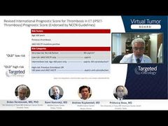
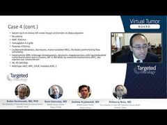
Then we get into our nondriver mutations. As we know, these are referred to as the HMR, or high molecular risk mutations, as Rami showed earlier when discussing the MIPSS70 [Mutation-enhanced International Prognostic Score System]. But here you have ASXL1, EZH2, SRSF2, IDH1/2, which all have…an increased risk of leukemic transformation in primary myelofibrosis.
I just want to point out a couple of things that are very striking. If you look at this slide and you particularly focus on IDH and TP53, you see how less than 5% of patients in chronic phase MF have these mutations. But look at what happens when they are in blast phase. The IDH numbers actually have been quoted to be even higher in some studies—up to 30%, TP53 also close to 30%. These 2 become very prominent in the post-MPN AML phase or the blast phase, which is very different from what we see in the chronic phase.
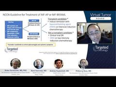
This slide just goes to show that there’s a lot that’s different between post-MPN AML’s genomic signature and that of de novo AML. As is very well known, FLT3 is mutated in up to 30% of patients with de novo AML. DNMT3A also tends to be very common. But here in post-MPN AML, you have a much lower incidence of FLT3 as well as DNMT3A, underscoring the point that this is a fundamentally different, biologically different disease.
Transcript edited for clarity.
Advertisement


Advertisement
Advertisement
Trending on Targeted Oncology - Immunotherapy, Biomarkers, and Cancer Pathways
1
Lung Cancer News Roundup: June 2026
2
Zoldonrasib Combos, Including With Daraxonrasib, Yield High Response Rates in PDAC
3
Speed Is the Story: How Sequencing the Human Genome Transformed Cancer Care
4
Examining the Strongest Options for Heavily Pretreated Myeloma After Bispecific Therapy
5